
 Same Day Clinic Hours
  (563)
(563) Imaging Center in Manchester
When it comes to taking a closer look inside your body, you will benefit from the highly skilled professionals in combination with advanced technology in Radiology at Regional Medical Center in Manchester, Iowa. This technology provides high quality imaging to help your doctor get a better diagnosis of your health concerns.
About Us
- A provider’s order is required for any diagnostic test to be performed. Bring the order with you (if it was an outside provider who ordered your test).
- Appointments are required for all imaging texts except x-rays.
- Have insurance card(s) ready to present.
- Radiology at RMC is completely digital. Meaning, images are taken and stored electronically, allowing Radiologists to access and read studies online. This process allows your healthcare provider to receive imaging reports in a more timely manner.
- Follow-up appointments are recommended for patients to schedule with their primary healthcare provider regarding their screening findings
Hours & Contact
Radiologists from Radiology Consultants of Iowa (Cedar Rapids) are also available Monday through Friday. After hour service and report interpretation is available via the Electronic Imaging Record.
563-927-7338 | Radiology Department
- Mon-Fri | 7AM-5:30PM
- Sat | 7AM-12PM
- Sun | Closed
Patient Parking & Entrance
Services Provided
- What is 3D Mammography?
- 3D Mammography is an x-ray of the breast tissue which is captured on specially designed digital detectors using low-dose radiation.
- The key role of mammography is to detect and identify breast cancer in its early stage. Studies have shown that when detected early, the chance for successful treatment of breast cancer is nearly 100%.
- Prepare for Exam:
- Dress in comfortable, two-piece outfit (you will need to undress from waist up)
- Do not use deodorants, powders or ointments around the breast and underarm area (this will affect quality of image)
- Do not schedule your mammogram just before or during your menstrual period (especially if you have breast pain during this time)
- Please inform the technologist if you have implants (additional imaging is required)
- During Exam:
- Breast tissue is compressed which helps to decrease radiation exposure and increase tissue detail
- Usually consists of two views of the breast (depending on breast size, additional imaging may be required)
- Diagnostic Imaging is used when abnormality is found during exam (breast mass, lump, nipple discharge, breast pain, skin thickening)
- Radiologist will interpret your images and a report will be sent to your healthcare provider
- Who should have a mammogram?
- The American Cancer Society recommends that at age 40 women should begin yearly mammograms. If you have a strong family history of breast cancer or other risk factors, your healthcare provider may recommend beginning earlier screening.
- You are required to have a provider order before scheduling your mammogram.
- What is bone density?
- Bone Densitometry is the test utilized for the evaluation of osteoporosis. Osteoporosis is the most common type of bone disease and most commonly associated with menopause.
- Prepare for Exam:
- Wear comfortable two-piece outfit without any metal buttons, buckles, or zippers
- During the Exam:
- You will lie on your back on a special table
- An overhead arm will scan above your body taking special readings of your bone density
- Your instructions will be to lie still while the images are being generated
- Measurements of your spine and hips are gathering and a computerized printout is then generated.
- The radiologist will interpret the images and report it to your healthcare provider
- What is a CT?
- CT (Computed Tomography or CAT) is an imaging test that assists in diagnosing a range of medical conditions
- CT imaging is useful because it can show several types of tissue such as lung, bone, soft tissue, and blood vessels with great clarity.
- Our Technology
- RMC’s 40-slice CT system brings state-of-the-art technology close to home. With this advanced technology, radiation exposure is kept at a minimum and automatically adjusts to each patient’s anatomy.
- If there is any chance of pregnancy, please tell your healthcare provider or the x-ray technologist.
- Prepare for Exam:
- Wear comfortable, loose-fitting clothing without snaps or zippers
- Some exams may require drinking a contrast liquid beforehand (these instructions will be relayed dependent on the specific exam you will be receiving)
- During the Exam:
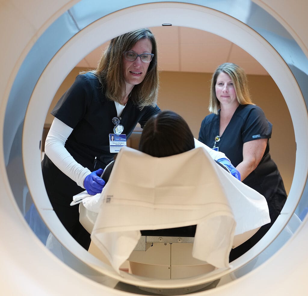
- Your instructions will be to lie on your back and the body part of interest slowly moved through the gantry (looks like a large donut, open at both ends)
- The x-ray system rotates around the gantry, taking detailed cross-sectional images
- Radiologist will interpret the images and report findings to your healthcare provider
- What is an MRI?
- An MRI uses a powerful magnet linked to a computer to create remarkably clear and detailed images of the internal organs and tissues without the use of radiation.
- Images are acquired of the specific body part from top-to-bottom, side-to-side, and front-to-back.
- Pacemakers or other implanted devices are NOT allowed in the Sheppard MRI Suite (stents are allowed, your trained MRI technician will review this prior to entering the MRI Suite)
- Prepare for Exam:
- Wear comfortable clothing with no metal buttons or zippers
- Most patients will be asked to put on a hospital gown
- All jewelry, hearing aids, removable dental work, and other metal objects will be removed prior to scanning
- Because of the nature of the machine, patients who undergo MRI may feel claustrophobic. If you are claustrophobic, please tell your healthcare provider so a light sedation may be given beforehand.
- During the Exam:
- A specially trained technologist will be observing you during your entire procedure and will be able to relay instructions during the test
- Earplugs or headphones are provided due to the noise made by the equipment during scanning
- A Radiologist will interpret your MRI exam and send a detailed report to your healthcare provider
- What is an Ultrasound?
- Ultrasound uses high-frequency sound waves that reflect off a body structure to form a special image.
- The echoes from the sound waves reflect back information regarding the size, structure, and uniformity of an organ; then are recorded; and displayed on a monitor screen.
- Ultrasound is happening in “real time” which means the screen displays what is happening in that moment.
- There is no radiation exposure with an ultrasound.
- There are a variety of reasons to have an ultrasound test ordered including obstetric, abdomen, gallbladder, pelvic, cartid, aorta and breast concerns
- Prepare for Exam:
- Your healthcare provider will give you specific instructions on how to prepare for your ultrasound exam
- Wear comfortable clothing, but you may be asked to change into a hospital gown
- During Exam:
- You will be asked to lie on the exam table
- A specially trained technologist will be performing the exam
- Lubricating gel is used to make a good contact between the ultrasound transducer (hand-held device that generate sound waves and sends it through the body) and the body part being evaluated
- Procedure is painless and usually takes 20-45 minutes to complete (dependent on your exam type)
- Radiologist will interpret your ultrasound procedure and send a report to your healthcare provider
- What is a Whole Body Composition Screening?
- Whole Body Composition Screening will help you learn about your whole body health, outlining the fat, lean muscle and bone using a scale of colors to represent each area.
- It’s a painless screening using a low-dose x-ray beam to take a picture of the inside of the body.
- This screening helps healthcare providers identify serious long-term health risks and can guide lifestyle changes to reduce risks
- Benefits of Screening:
- Able to see the areas of the body storing extra fat
- More accurate than BMI (body mass index)
- Monitors body fat loss and muscle growth from exercise
- Provides baseline for nutritional counseling and treatment for weight-related issues
- Maximizes performance of athletes
- Prepare for Screening:
- Wear clothing with no zippers, buttons or metallic parts (hospital gown available)
- Remove all jewelry, hairpins, glasses and other metallic objects
- During Exam:
- Plan approximately 30 minutes for the entire procedure including registration
- The scan itself will last 6 minutes
- Park in Lot 2, Entrance E
- Register at West Registration (Emergency/Surgery Registration)
- $100 payment is due at the time of service